Research Area
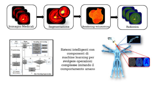
Non-invasive imaging techniques: Positron Emission Tomography (PET), Computerized Tomography (CT) and Magnetic Resonance (MR)
Big data, Radiomics and Artificial Intelligence in Clinical Health Care Applications
Processing, quantification, and correction methods for ex-vivo and in-vivo medical images.
National collaborations
Nuclear Medicine Department, Giglio Hospital, Cefalù, Italy
Nuclear Medicine Department, Cannizzaro Hospital, Catania, Italy
Nuclear Medicine Department, Messina University Hospital, Messina, Italy
Radiology Unit, Palermo University Hospital, Messina, Italy
Radiology Unit, Catania University Hospital, Messina, Italy
Ri.MED Foundation, Palermo
International collaborations
Prof. Anthony Yezzi, Laboratory of Computational Computer Vision (LCCV), School of Electrical and Computer Engineering, Georgia Institute of Technology, Atlanta, USA
Dr. Samuel Bignardi, Laboratory of Computational Computer Vision (LCCV), School of Electrical and Computer Engineering, Georgia Institute of Technology, Atlanta, USA
References
- Comelli A, Stefano A, Coronnello C, Russo G, Vernuccio F, Cannella R, Selvaggio G, Lagalla R, and Barone S. Radiomics: a New Biomedical Workflow to Create a Predictive Model. Communications in Computer and Information Science. Springer, Cham, 2020. In press.
-
- Stefano A, Comelli A, Bravata V, Barone S, Daskalovski I, Savoca G, et al. A preliminary PET radiomics study of brain metastases using a fully automatic segmentation method. BMC Bioinformatics, 2020. In press.
-
- Stefano A, Gioè M, Russo G, Palmucci S, Torrisi SE, Bignardi S, et al. Performance of Radiomics Features in the Quantification of Idiopathic Pulmonary Fibrosis from HRCT. Diagnostics 2020, Vol 10, Page 306 2020;10:306. https://doi.org/10.3390/DIAGNOSTICS10050306.
-
- Comelli A, Stefano A*, Russo G, Bignardi S, Sabini MG, Petrucci G, et al. K-nearest neighbor driving active contours to delineate biological tumor volumes. Eng Appl Artif Intell 2019;81:133–44. https://doi.org/10.1016/j.engappai.2019.02.005.
-
- Comelli A, Stefano A*, Bignardi S, Russo G, Sabini MG, Ippolito M, et al. Active contour algorithm with discriminant analysis for delineating tumors in positron emission tomography. Artif Intell Med 2019;94:67–78 https://doi.org/10.1016/j.artmed.2019.01.002.
-
- Comelli A, Stefano A*, Russo G, Sabini MG, Ippolito M, Bignardi S, et al. A smart and operator independent system to delineate tumours in Positron Emission Tomography scans. Comput Biol Med 2018;102:1–15. https://doi.org/10.1016/j.compbiomed.2018.09.002.
-
- Raccagni I, Belloli S, Valtorta S, Stefano A, Presotto L, Pascali C, et al. [18F]FDG and [18F]FLT PET for the evaluation of response to neo-adjuvant chemotherapy in a model of triple negative breast cancer. PLoS One 2018;13. https://doi.org/10.1371/journal.pone.0197754.
-
- D`Urso D, Stefano A, Romano A, Russo G, Cosentino S, Fallanca F, et al. Analysis of Metabolic Parameters Coming from Basal and Interim PET in Hodgkin Lymphoma. Curr Med Imaging Rev 2017;14:533–44. https://doi.org/10.2174/1573405613666170331110119.
-
- Rundo L, Stefano A, Militello C, Russo G, Sabini MG, D’Arrigo C, et al. A fully automatic approach for multimodal PET and MR image segmentation in gamma knife treatment planning. Comput Methods Programs Biomed 2017;144:77–96. https://doi.org/10.1016/j.cmpb.2017.03.011.
-
- Gallivanone F, Stefano A, Grosso E, Canevari C, Gianolli L, Messa C, et al. PVE correction in PET-CT whole-body oncological studies from PVE-affected images. IEEE Trans Nucl Sci 2011;58:736–47.


